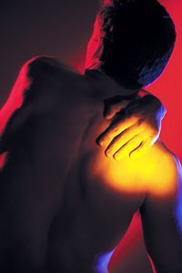
Shoulder Pain: Practical Tips for Examination and Treatment
By Kevin M. Wong, DC
I have been fortunate thus far in my chiropractic career to have been exposed to many different types of patients with a variety of ailments conducive to chiropractic care. Although I will always love my roots in spinal adjusting, I really get a charge out of doing extremity work. Let’s focus on one of the most common problem areas people come to see us for: the shoulder.
To set the stage for a moment, we need to get a realistic visualization of how our patients’ (and our) shoulders can become painful in the first place. Just a few of the many causes include poor sitting or desk posture, injuries, sleeping on your side, and other activities during which your arms are in front of you (driving, sewing, gardening, etc.). Almost any arm activity can create the potential for shoulder problems. Since we stress and overstress our shoulders in our typical daily living, shoulder problems are a natural consequence.
Take a second and palpate the ball and socket joint of each of your shoulders. What do you feel? Any tenderness or pain? Any asymmetries in how the shoulders are hanging? In most of my patient population, I can palpate the anterior part of the humerus and find tight or hypertonic muscles. Yes, the anterior deltoid, biceps tendon and even the pectoralis muscles in the front tend to be tighter and more sore than the posterolateral areas. Do they feel sore to you?
General Analysis
 Remember all of your shoulder joints: glenohumeral (GH), acromioclavicular (AC), sternoclavicular (SC), scapulothoracic and rib joints (costovertebral, costotransverse and costosternal). Also recall that you have a battery of orthopedic and neurologic tests available to you when assessing the shoulder. You are not expected to have all of them memorized, so have references available to use when you need to be reminded. These tests can add critical information to your fact finding and treatment plan.
As we are practice, our diagnostic, palpation and kinesthetic skills improve. The more you put your hands on people, the better you get. You may know exactly what is wrong with your patient’s shoulder from the history itself, but follow through with the rest of your exam.
Now let’s talk about the shoulder joints one by one and how they generally misalign. Keep in mind that this is not intended to be a comprehensive look at every aspect of the shoulders. Let’s keep the focus strictly on how most patients typically present with shoulder problems in your office.
Shoulder Joint Misalignment
GH joint: A multi-axial synovial ball-and-socket joint, the GH joint is the articulation between the glenoid fossa of the scapula (shoulder blade) and the head of the humerus (upper arm bone). Due to its shallow socket, the presence of a fibrocartilaginous labrum aids in support. We also see the rotator-cuff muscles attaching on the head of the humerus. The GH joint is the most mobile and least stable joint in the human body.
Our patients understand the GH as the major “shoulder joint” and it is the primary one that gets attention. In our society, we do most of our activities in front of our bodies with the shoulders rounded. As a result, patients often present with an anterior-inferior (AI) humerus.
When the patient is sitting or lying on their back, you can see one of the humerus bones protruding higher than the other, non-symptomatic side. Even touching the front of the higher humerus can be painful. Of course, the humerus can also move posterior or lateral, but those tend to be more of a traumatic circumstance. A lion’s share of your patients will have the AI shoulder.
AC joint: This joint is the junction between the acromion (part of the scapula that forms the highest point of the shoulder) and the distal clavicle. The AC joint allows the ability to raise the arm above the head. It is a gliding synovial joint that acts as a pivot point for movement of the scapula, resulting in a greater degree of arm rotation.
Patients refer to this joint at the point or tip of their shoulder. In this area, the distal clavicle will misalign in a superior direction. This movement is not like a shoulder separation, which also occurs at this joint. Shoulder separations involve ligamentous damage or tears and could, in severe cases, require surgery to repair.1
A good thing to remember is that when someone has suffered a prior fracture of the distal clavicle, it will be obvious when you look at them, as the bones rarely heal straight. Look for that bony callous or deformity; it is hard to miss.
SC joint: The sternoclavicular is a synovial joint composed of two portions separated by an articular disc. The joint is made up of the sternal end of the clavicle, the upper and lateral part of the breastbone, and the cartilage of the first rib.
Ligaments that help attach the proximal clavicle to the sternum are very strong here. We rarely find fractures in this region. We do get misalignments of the proximal clavicle, most commonly moving superior/anterior/medial. Be especially careful when you palpatethis joint. Not only can they be particularly tender, but you are also getting close to the chest area. Take the appropriate precautions when working with female patients.
Scapulothoracic joint: The scapulothoracic joint is not a true joint, in the sense that it has no capsule or ligamentous attachments. It is more commonly referred to as an articulation. It is formed between the anterior scapula and the posterior thoracic rib cage (ribs 2-7).
The scapula’s attachment to the skeleton is musculotendinous in nature, formed by the trapezius and serratus muscles.
Its gliding movement patterns consist of elevation/depression, retraction/protraction, and superior/inferior rotation. Often, we find that the shoulder blades do not “flare” equally. Fibrous adhesions can occur following a shoulder injury, especially if the joint has been immobilized for a long period of time. This impairs movement of the shoulder.
Costovertebral, costotransverse, and costosternal (rib) joints: Costotransverse joints involve the facets of the tubercles of ribs 1-10, forming joints with the corresponding thoracic vertebrae. These are synovial joints. This articulation is present in all but the 11th and 12th ribs. Ribs 1-10 have two joints in close proximity posteriorly: thecostovertebral joints and the costotranseverse joints. The costosternal joints are those involving the cartilages of the true ribs with the sternum. These are considered arthrodial joints, with the exception of the first joint, where the cartilage connects directly with the sternum (synarthrodial).
Ribs are present to help protect the vital organs and assist with inspiration / expiration. Pain felt from ribs out of alignment can be some of the sharpest, most intense pain felt anywhere. Often, sharp rib pain can shoot through the chest and be mistaken for angina. It causes a lot of people to run to the emergency room in a panic.
Treating Shoulder Problems
So, how do you know when to check the shoulder? Beyond the obvious shoulder pain symptoms (the patient is pointing directly at it), you might look at the shoulder when you see any of the following:
- Pain in the trapezius, especially the region just above the scapula
- Neck pain that can move into the skull
- Referral of pain or isolated pain in the lateral or anterior deltoid
- Popping or clicking of the shoulder during AROM or PROM
- Weakness with muscle or strength testing of the GH muscles
- Patients pointing to rib joints as sites of pain. Ribs are often out due to shoulder misalignments
- Chronic/persistent C/T pain that is not resolving with spinal adjustments
When it comes to adjusting the shoulder joints, keep in mind what we discussed previously about the directions they tend to misalign.2 We have a wide buffet of adjusting maneuvers and techniques available to us, so here are a few pointers, including manual adjusting techniques, drop-table adjusting and instrument adjusting.
Manual adjusting techniques: These can be very effective, but also very painful. Watch out how much stress and force you apply. Whichever position you place the patient in, make sure to feel for the movements you are trying to achieve for each of the joints. Getting a “pop” should not always be the objective.
Drop-table adjusting: Supine drop adjusting is another effective way to address these joints. Remember that the tension set on the table must be light. The joints can be very tender, so watch your depth of pressure with your fingers when you set up and engage the drop piece. The thumbs or the thenar/hypothenar contacts both work well and are generally comfortable for patients.
Instrument adjusting is also great for addressing the shoulder joints. For some patients, especially children, healthy adults in acute pain and the elderly, this is actually the preferred way to start moving the bones. You can always transition them into other light-force techniques, like drop table. Don’t be afraid to stick with instrument adjusting if the patient likes it and is responding well.
Chiropractic adjustments and treatments are proven to relieve the pain associated with many common shoulder injuries. “There is fair evidence for the treatment of a variety of common rotator- cuff disorders, shoulder disorders, adhesive capsulitis, and soft-tissue disorders using MMT [manual and manipulative therapy] to the shoulder, shoulder girdle, and/or the full kinetic chain (FKC) combined with or without exercise and/or multimodal therapy.”3 The most important aspect is to recognize and address all of the shoulder joints during your assessment. Adjust each individual joint, taking into consideration which direction you feel the bones have moved, and see how successful the results are. The goal is simple: provide the most efficient, effective patient care.
References
- Mazzocca AD, Arciero RA, Bicos J. Evaluation and treatment of acromioclavicular joint injuries. Am J Sports Med, 2007 Feb;35(2):316-29.
- Hudson VJ. Evaluation, diagnosis, and treatment of shoulder injuries in athletes.Clin Sports Med, 2010 Jan;29(1):19-32.
- Brantingham JW, Cassa TK, Bonnefin D, et al. Manipulative therapy for shoulder pain and disorders: expansion of a systematic review. J Manip Physiol Ther, 2011 Jun;34(5):314-46.
Shoulder Pain: Practical Tips for Examination and Treatment